Table of Contents
PART II
BIOLOGY AND EXERCISE PHYSIOLOGY
WIESŁAW CHWAŁA, WANDA FORCZEK
Department of Anthropomotorics, Academy of Physical Education in Krakow, Poland
Correspondence should be addressed to: Wiesław Chwała, Department of Antropomotoricity, Biokinetics Workshop, Academy of Physical Education in Krakow, 31-571 Krakow Poland, 
Table of Contents
Key words: humangait, Vicon system, angular changes, extremities.
Perry suggests that during walking the body functionally divides itself into two units: passenger and locomotor. The former consists of the head, neck, trunk and arms (named HAT by Elftman), which is a structure on top of the locomotor apparatus, responsible for its own postural integrity. The latter is formed by the two lower limbs and pelvis. Its main task is to carry the body forward (including the HAT). Besides, the locomotor is responsible for propulsion, stance stability, and shock absorption. The aim of this study was to assess the values of angular changes in reference to the HAT parts in all three planes of movement in able-bodied subjects during gait with natural velocity. The study was based on a sample of 40 healthy volunteers aged 20-25 years. In order to avoid any disturbances of the correct results the inclusion criteria aimed at keeping in the sample only subjects without any locomotor disorders, i.e. no history of orthopaedic or neurosensory disorders. A three-dimensional Vicon system was used during the spatial movement analysis.
Walking is one of the most common and complex of all human activities. Perry suggests that during walking the body functionally divides itself into two units, passenger and locomotor. The first unit consists of the head, neck, trunk and arms (named the HAT by Elftman) which is structure on top of the locomotor apparatus, responsible for its own postural integrity. The two lower limbs and pelvis form the locomotor system responsible for carrying the passenger unit forward. Other locomotor functions are propulsion, stance stability and shock absorption. Lower limbs and pelvis movements during the gait have been subject to numerous studies before [1, 3, 5, 7, 8].
The aim of this study is to analyze and assess the values of kinematic parameters of the gait with natural velocity, in reference to the biokinematic chain of the upper body. Natural velocity is defined as an individual’s normal walking speed. Its average value in this study was 1.41±0.16m/s. As far as movement kinematics of a subject during locomotion is concerned, it is necessary to intensify the study of biomechanical values of upper body parameters, because research in this field is far from complete and all the three planes are rarely included. Such studies can be applied not only as an evaluation of the change ranges, changes directions of parameters, and disparity of results in a normal group of people, but as a biomechanical pattern of movement, also if we want to identify a dysfunction of the body structure resulting from different sport injures. Besides, this analysis can contribute to better understanding of the gait mechanics and help to undertake proper clinical treatment in case of any pathology.
The study was carried out in 2003 during a Biomechanics Workshop in the Department of Anthropomotorics at the Academy of Physical Education in Krakow. The study was based on a sample of 40 volunteers aged 20-25 years, using the Vicon optoelectronic system with five cameras. With this system it was possible to establish three-dimensional trajectories of markers fixed on the subjects' skin. The cameras were linearized and the whole system was calibrated according to manufacturer's instructions.
Inclusion criteria aimed at keeping in the sample only subjects without any locomotor disorders, i.e. with no history of orthopedic or neurosensory disorders that can affect the results of the study.
For each subject 12 gait cycles were recorded with normal stabilized velocity. On the basis of the analysis of 480 normalized walking cycles the mean values of biomechanical parameters were established that characterized locomotion of the trajectories [6]. In the description of the results a division of the gait into eight phases was applied, which was suggested by Perry in the Los Amigos Medical Center (RLA). These were: 1. Initial Contact– IC (0%); 2. Loading Response– LR (0-10%); 3. Midstance – MST(10-30%); 4. Terminal Stance–TST (30-50%); 5. Preswing– PSW (50-60%); 6. Initial Swing– ISW (60-70%); 7. Midswing–MSW (70-85%); and 8. Terminal Swing–TSW (85-100%).
Gait phases were given in per cent gait cycle, i.e. from the heel strike of an analyzed limb to the next heel strike of the ipsilateral limb. Also another division of human gait is used, where the cycling character of the gait (phasing) is the main criterion [3]. It consists of two phases: a) single support (floor contact with one leg), with the time of support approximately 0.53 s; and b) double support (both legs touching the ground), with the time of support approximately 0.15 s. The data connected with gait refers to the frequency of 90 steps/min [3]. However, gait is usually described in reference to the activity of one extremity. The gait cycle is divided into two phases: stance and swing, and two periods of double support (Tab. 1).
Table 1. Traditional division of the gait

The term ‘double support’ refers to the two intervals in a gait cycle, in which body weight is transported from one foot to the other. Both feet are in contact with the supporting surface at the same time [8].
VICON system characteristics
The study was carried out using the Vicon system which makes it possible to record and analyze movement in a three-dimensional space. This study is not invasive. It involves passive markers fixed directly on the subjects' skin that reflect localization of the characteristic points, and joints’ axes work using passive markers. The markers in the semi-spherical form are made of a semi-reflecting material. Vicon enables to establish three-dimensional trajectories of markers in the form of points as well as their dimensional changes.
This system consists of five video cameras, a datastation (Fig. 1) and infra-red projectors. The speed of image acquisition depends on the type of camera and its localization which determines the speed of the analyzed movements. The cameras work with the standard frequency of 120 Hz.
The Vicon datastation consists of a specialized computer that collects and analyses information recorded by the cameras. Then, it sends the information to a PC with applications for analysis of the research material.
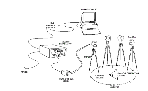
Figure 1. The measurement line scheme of Vicon system. The source: Vicon Software Manual.
At the beginning the study requires preparation of the measurement area and system. The measurement area is a place within the cameras’ field. During the human locomotion study an area of 8 meters in length was used, which allowed recording four gait cycles. Then the whole system was calibrated according to manufacturer's instructions. The markers indicated particular body segments – hence they had mainly been placed in the joints axes – within a given distance from the symmetry joint centre and characteristic points on the head, chest, and pelvis, which allowed reflection of these body segments in space, and measurement of the relevant parameters, e.g. chest or pelvis width (Fig. 2). The accurate location of the markers enabled a proper description of the centres of joint symmetry [5].
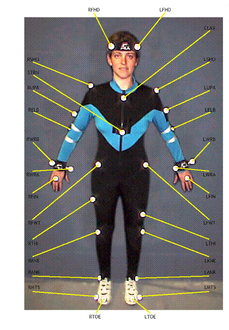
Figure 2. Set of the markers for the body – anterior view The source: Vicon Software Manual.
The movement trials were followed by measurements of the corresponding anthropometric parameters, considering the somatic body build, which were fed into the computer afterwards. When anthropometric data were matched with the GOLEM in BODY BUILDER mathematic model, the actual location and the subject’s body movement were graphically displayed. As a result, a set of kinematic parameters was achieved, consisting of spatio-temporal parameters, angular changes, velocities and angular accelerators within joints, anthropometric points’ trajectories in all three planes (sagittal, coronal and horizontal), and finally length changes of the chosen muscles during a normalized gait cycle.
Shoulder angles
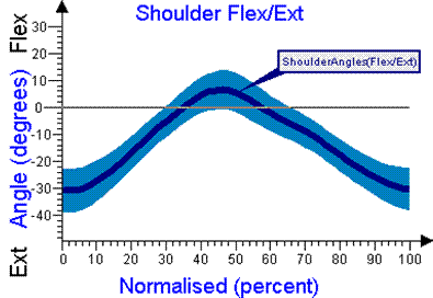
Shoulder angle changes are related to the shoulder movements according to the trunk in three planes. Figure 3A presents a two-directional shoulder movement in the sagittal plane in the following way: flexion is seen from IC to the end of TST; and from the beginning of ISW to the end of the gait cycle (TSW) extension. The shoulder stays in the position of maximum extension (32°) at the onset of stance, and maximum shoulder flexion is reached near the end of the terminal stance. Slightly later the elbow completes its flexor action. As far as this joint is concerned the range of angle changes is approximately 39±8°. We can notice two neutral positions of the shoulder during the TST and ISW phases.
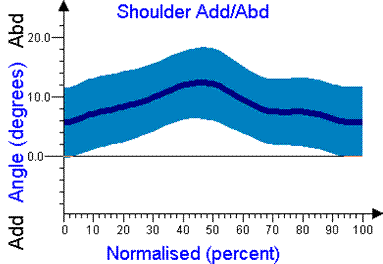
Shoulder movement in the coronal plane (Fig. 3.B) displays characteristic angular changes for normal gait in relation to abduction and adduction movement. The movement in this plane is smooth and characterized by a larger disparity of individual results than in the sagittal plane. This is an indication of a great variation in the angular range in subjects. What is important is that the entire movement in the coronal plane takes place in abduction arrangement according to the joint axis. From the beginning of the foot flat until the end of the TST position the shoulder abducts. The range of movements (Fig. 3.C) varies significantly among the subjects. This is an indication of a great disparity at the onset position of the shoulder and its rotation in the transverse plane. After holding this position of peak flexion momentarily, the shoulder extends then through the swing phases.
A
B
C
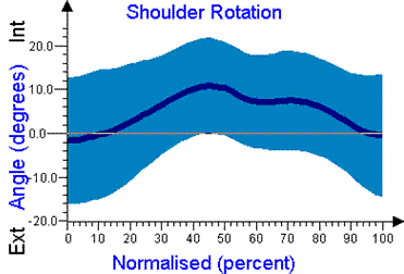
Figure 3. Angular changes in the shoulder joint. A. In the sagittal plane (flexion/extension) in normalized gait cycle. B. In the coronal plane (abduction/adduction) in normalized gait cycle. C. In the transverse plane (internal /external rotation) in a normalized gait cycle.
Elbow angles
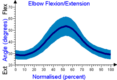
All the time during walking the elbow stays in the flexion position (Fig. 4.A). Moving in the same direction as the shoulder, the elbow also goes through a corresponding arc of flexion and extension during each stride. At the onset of the stance it flexes to a position of 30°, however, never extending beyond 20° flexion. As a result, maximum flexion (56°) is achieved by the time of the contralateral foot flat.
Contralateral foot flat at the onset of pre-swing stimulates both the shoulder and elbow to reverse their motion toward extension. This motion continues through the swing period. The elbow reaches its maximally extended position of 20° flexion by the mid swing, while the shoulder continues extending until its final posture of 7° is achieved as the ipsilateral heel contacts the floor once again. The total range of motion in this joint is 28±4°.
Flexion and extension movements in the sagittal plane are accompanied by rotation movements in the horizontal plane (Fig. 4.B). Their range is approximately 7°, whereas the entire movement stays in interior rotation of the forearm.
A
B
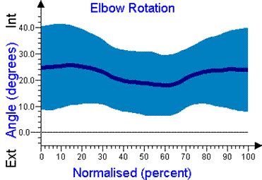
Figure 4. Angular changes in the elbow joint. A. In sagittal plane (internal /external rotation) in normalized gait cycle. B. in transverse plane (internal /external rotation) in normalized gait cycle.
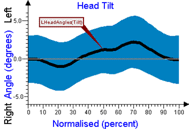
Head angles
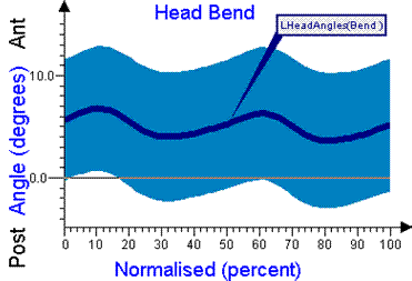
Head angles relate to the body of walking subject, i.e. “Anatomy”. The head is bent anterior through the entire range of mean angular values. Its range of motion (just about 3°) is characterized by a significant disparity of the results in individuals, as shown by the relatively large shaded area. Despite this variability among the subjects, the patterns of gentle changes of head position can be noticed. The head angles oscillate between 1.5 and 4.5° showing two peak deflections within each walking cycle (Fig. 5A).
Head movements in the coronal plane feature small angular changes (3°). At the onset of the stance the head is near the neutral position (Fig. 5B). In the MST the first movement we can observe is towards the long axis of the body (approximately 1.5), and then the second one, which lasts until the end of the ISW, is lateral tilting of the head. At the end of the swing phase (TSW) the head returns to the neutral position.
The average total angular range in the transverse plane is approximately 10°. At IC the head position near to neutral can be observed (Fig. 5C). The greatest movement of head rotation is connected with TST, the PSW phase, and the end of the swing phase (TSW). During ISW and partly in MSW we have found a very similar pattern of angular changes as during the phases at the onset of the stance (the range of rotation reaches approximately 3°). Then the head rotates to the left to a position of 8° rotation.
A
B
C
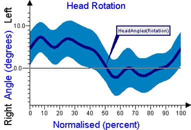
Figure 5. Angular changes in the head. A. In sagittal plane (anterior/posterior bending) in normalized gait cycle. B. In coronal plane (right/left tilting) in normalized gait cycle. C. In transverse plane (right/left rotation) in normalized gait cycle.
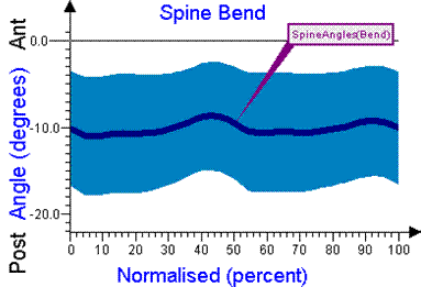
Spine angles
Spine angles (whole lumbar segment) are recorded in relation to the pelvis. Lumbar spine movements according to the pelvis in the sagittal plane feature a small range of angular changes, approximately 3°. Simultaneously, this part of the spine is in a flexed position according to the pelvis during the entire normalized gait cycle. We have found significant changes in the LR, TST and TSW phases. Figure 6A presents two peaks of extension (minimal value of flexion) at 40% and 90% of the gait cycle. The disparity of the individual flexion lumbar spine position according to the pelvis should be emphasised. It results from the variability of the physiological curve of the analyzed spine unit.
Figure 6B shows the trajectory of the lumbar spine in the coronal plane, which features three basic angular changes. At IC this segment is in neutral position, and then in LR it bends to the left according to the vertical pelvis axis, to the position of 6°. From the onset of MST to the end of the stance phase in PSW a spine movement to the left in the range of 12° can be noticed. During the first part of TST the lumbar spine is in neutral position according to the pelvis, which goes through a 10% gait cycle. After toe-off, during ISW and MSW, the spine returns to the neutral position. At the end of the swing phase TSW the lumbar spine does not change its position in reference to the pelvis in the coronal plane.
The range of angular changes during rotation is significant (approximately l2°) (Fig. 6C). The largest rotation angle (6°) is in IC, then there is a movement in the right-side rotation, which achieves maximum at the end of TST (6°). In PSW left rotation movement begins and continues until the end of the gait cycle, where it reaches the value of 6°. The shaded area, which presents a disparity of the results in the subjects, oscillates between 6-8°.
A
B

C
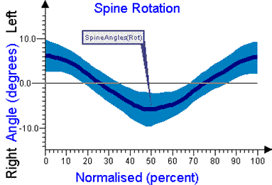
Figure 6. Angular changes in the lumbar spine. A. In sagittal plane (anterior/posterior bending) in normalized gait cycle. B. In coronal plane (right/left tilting) in normalized gait cycle. C. In transverse plane (right/left rotation) in normalized gait cycle.
If we take into consideration the great role of the passenger unit (HAT) in locomotion, it seems to be a key issue, if we want to understand the reciprocal influence on the particular segments of the body in relation to the entire biomechanism movement [6, 10]. Dysfunctions which result from pathological work of the lower extremities or pelvis are always reflected in compensatory work of the upper body unit. Besides the range of angular changes in all three planes that accompanies HAT can remain uninfluenced on the lower limb work [11, 12].
Our considerations in this paper have focused on studying and better understanding of the work mechanism of the upper limbs, trunk and head in the able-bodied subjects during gait with natural velocity. Subjects’ gait was analyzed by means of kinematic parameters of the gait. Angular changes of the shoulder and elbow joints, head and lumbar spine movements in all three planes: sagittal, coronal and transverse, were taken into account.
As there have not been many investigations in this field, the results of this study can serve as interesting characteristics of gait in the population of healthy people.
In the study by Bober [4] no accurate data in reference to angular changes in all upper limb joints can be found. He just noticed the reciprocal action of upper and lower extremities, where integrated movements of the pelvic and shoulder girdle reflect particular phases of the gait. Although upper limb rotation movement is not the main condition of the proper gait, upper extremities; however, it plays an important role in the total gait pattern. Based on the preliminary observation of the rotation patterns of the upper limbs, Murray [6, 7] claims they are the most variable gait components. It has also been confirmed by our own studies.
Shoulder angles present a two-directional shoulder movement in all three planes. The change of movement direction becomes visible at the end of the TST phase. The shoulder flexes from maximum extension at the onset with reciprocal abduction and interior rotation. In the second part of the cycle the shoulder extends through the swing phase and diminishes abduction and interior rotation (returning to neutral position in the transverse plane).
Phasing of arm motion during walking is quite distinct. At initial contact the ipsilateral arm is maximally extended at both the shoulder and elbow. Following a brief delay, the shoulder flexes progressively. There is a greater delay at the onset of elbow flexion, which may relate to the maximally extended position of 20° flexion. Movement at the elbow toward greater flexion begins in mid stance. Moving in the same direction as the shoulder, the elbow also goes through an equivalent arc of flexion and extension during each stride. All the time during walking, the elbow stays at the flexion position. Contralateral foot flat at the onset of pre-swing stimulates both the shoulder and the elbow to reverse their motion toward extension. This motion continues through the swing period.
Cappozzo [1] described the head movement trajectories in all three planes on the basis of other similar studies. The results of our investigation reflect his conclusions.
The head is bent anterior through the whole range of mean angular values. Head movement in the coronal plane features small angular change. These are only small oscillate movements. At the onset of the stance the head is near the neutral position. The average total angular range in the transverse plane is larger than in the sagittal plane. The largest head rotation movement is connected with the TST and PSW phase, and at the end of swing phase. During ISW and partly in MSW we have found very similar angular changes to the phases at the onset of the stance.
The lumbar spine segment is flexed in relation to the pelvis during the entire normalized walking cycle. In relation to the pelvis this part of the spine features a small range of angular changes in the sagittal plane. Significant differentiation of the individuals’ flexion alignment results from differentiation within physiological curves of the analyzed part. A large range of disparity (3l-17.5-) in subjects can be noticed.
In the coronal plane we have found movement towards the vertical axes of the pelvis and resting of the stance phase accompanies a movement to the right from the pelvis axes. Simultaneously, these movements are supported by a rotation to the left. In the swing phase we can see a significant movement of the lumbar segment toward the vertical pelvis axes in the sagittal plane with the right-side rotation of the entire segment. In the sagittal plane we can notice a smooth movement toward spine extension at the end of the swing phase.
Allard, P., Cappozzo, A., Lundberg, A., Vaughan, C.L., Three-dimensional Analysis of Human Locomotion, Wiley & Sons New York 1997.
Allard, P., Lachance, R., Aissoui, R., Sadeghi, H., Duhaime, M., Able-bodied Gait in Men and Women, (in:) Three-dimensional Analysis of Human Locomotion. 1997.
Allard, P., Stokes, I.A.F., Blanchi, J. P., Three-dimensional Analysis of Human Movement, Human Kinetics, New York 1995.
Bober, T., Biomechanics of gait and running, AWF Wrocław 1985.
Hof, A., Scaling gait data to body size, Gait Posture, 1996, 4, pp. 222-223.
Murray, M.P., Drought, A.B., Kory, R.C., Walking Patterns of Normal Men, ”The Journal of Bone and Joint Surgery”, 1964, pp. 335-360.
Murray, M.P., Kory, R.C, Sepic, S.B., Walking Patterns of Normal Women, “Archives of Physical Medicine & Rehabilitation”, 1970, pp. 637- 650.
O’Sullivan, S.B., Schmitz, T.J., Physical Rehabilitation: Assessment and Treatment, Philadelphia, pp. 195-218.
Ounpuu, S., Winter, D., Bilateral electromyographical analysis of the lower limbs during walking in normal adults, “Electroencephalo. Clinical Neurophysiology”, 72. 1989.
Perry, J., Gait analysis, Thorofare, SLACK 1992.
Saunders, M., Inman, V.T., Eberhard H.D., The major determinants in normal and pathological gait, “Journal of Bone and Joint Surgery”, 1953, pp. 543-558.
Vaughan, C.L., Davis, B.L., O’Connor, J.C., Dynamics of Human Gait, Kiboho Publishers Cape Town South Africa 1999.
Winter, D., Biomechanics and motor control of human movement. Wiley & Sons, New York, 1990.